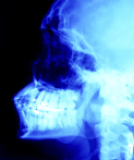
 Dentists use x-rays which utilize ionizing radiation, in order to obtain information regarding the health of the tooth, the gums and the bone tissue which surrounds the tooth. Although these types of x-rays don’t tend to be absorbed by your tongue and gums, they are absorbed by your teeth, bones and surrounding hard tissue. The problem with this is that ionizing radiation is considered the primary environmental cause for the most common type of brain tumor in the United States– meningioma. Although you are exposed to ionizing radiation through natural sources, dental x-rays are the biggest source of exposure to this tumor causing radiation. Recently scientists set out to examine just how dangerous these dental x-rays are and the results, which were published in the April 2012 issue of the journal Cancer, were shocking.
The study was led by Elizabeth Claus, MD, PhD, of the Yale University School of Medicine in New Haven and Brigham and Women’s Hospital in Boston. Claus and her colleagues examined data obtained between May 1, 2006 and April 28, 2011. The data included 1,433 meningioma patients between the ages of 20 and 79, who resided in multiple American States. In addition, the researchers also examined data from a control group of 1,350 individuals who did not have meningioma.
Advertisement
The researchers found that the patients diagnosed with meningioma were more than doubly likely than their healthy counterparts to have had a bitewing exam, which uses an x-ray film, kept in place by the patient’s teeth and gums. Furthermore, those patients who had received a bitewing exam yearly were 1.4 to 1.9 times more likely to develop meningioma than those who did not.
How the Dentist Should Keep You Informed
Another type of ionizing radiation procedure that many a dentist will use is the panorex x-ray, which gives the dentist a complete picture of each tooth. The researchers found that individuals, whose dentist had given them a panorex x-ray before the age of 10, had almost a 5-fold increased risk for developing meningioma. In addition, individuals who received this x-ray on a yearly bases had a 2.7 to 3 times greater likelihood for developing meningioma.
The type of radiation that a patient is exposed to when their dentist gives them an x-ray, does not only increase brain tumor risk, it has also been associated with an increased risk for leukemia, thyroid, breast, lung, colon, bladder, ovary and stomach cancer. According to the National Institute of Health, even low doses of ionizing radiation can cause direct damage to a person’s cells and DNA, and if that is in fact true, then it means that ionizing radiation can contribute to all types of cancer.
Currently, the American Dental Association recommends that healthy children have one x-ray every 1-2 years, that teens have one x-ray every 1.5-3 years, and that adults have one x-ray every 2-3 years. More worrying is the fact that some dentists take this recommendation a step further and perform bitewing x-rays every 6 months, even on patients with outstanding oral health.
Given the associated risks with dental x-rays, perhaps it is time for the dentist community to re-valuate this recommendation. While completely eliminating the use of dental x-rays is a highly unlikely proposition; performing them based on necessity as opposed to a preplanned schedule, could help to vastly decrease the patient’s lifetime exposure to the radiation. According to Dr. Nicholas Dello Russo, an instructor at the Harvard University School of Dental Medicine, dentists should rely on visual exams of the mouth, and patients who display healthy teeth and gums, and who are not experiencing any problems should consider forgoing these yearly exams.

