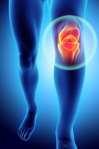
There are four different plica synovial folds that can be in the human knee; however, there is only one that causes trouble. It is the medial plica, which attaches to the lower end of the patella (knee). It is believed that anywhere between 50 to 70 percent of us have a medial plica, yet it never causes us any problems. Unfortunately though, some people do get medial plica syndrome. Medical researchers suggest that plica is remnants of embryonic connective tissue that just didn’t fully resorb while the fetus was developing.
We have to imagine that the knee joint is a sleeve of tissue. This sleeve is made up of synovial tissue, which is a thin, slippery material lining all joints. The synovial sleeve of tissue has folds that allow movement of the joint without any restrictions. Plica syndrome or synovial plica syndrome inflames the knee. Your plica can catch during knee straightening and bending, when you experience blunt trauma, have altered knee motions, or have knee issues, such as a torn meniscus. Plica rarely occurs on its own, usually there is another knee condition at play.
We mentioned meniscal injury, but a person who is suffering from knee plica syndrome could also have a condition known as patellar tendonitis. Some people with plica syndrome of the knee suffer from something called Osgood-Schlatter’s Disease. OSD is a growth-related problem that is mostly seen in active young boys. It can occur during a growth spurt.
Causes and complications of plica syndrome
As mentioned, knee plica syndrome has been linked to embryonic development. During the development phase, the knee begins as a division of synovial membranes. In fact, they are three separate compartments. However, by the third to fourth month of fetal development, the membranes are resorbed and the knee transforms into a single chamber. These remnants are called synovial plicae. We can’t say for sure why symptomatic plica occurs in some individuals, but there are theories about causes of plica syndrome.
Inflammation, a single blunt trauma, loose bodies, meniscal tears, osteochontritis dissecans, which is a joint disorder where cracks seem to form in the cartilage and subcondral bone, are suspected causes. For many people, stressing or overusing the knee can cause plica syndrome. Those who exercise the knee frequently are at risk of symptomatic plica. For instance, runners, bikers, and those who use stair-climbing machines on a regular basis are more prone to plica syndrome.
Symptoms of plica syndrome
There are some people who experience injuries or even multiple surgeries near the medial area of the knee. These situations can lead to a thickening of the synovial plica. Think of it as an excessive amount of fibrous tissue. This very thick, fibrotic material has the potential to catch over the femur, which is the thighbone and happens to be the longest bone in our bodies.
Here are some of the common plica syndrome symptoms:
- Knee pain
- Audible clicking or snapping during knee motion
- Tender plica when touched
- Pain associated with activities, including stairs, squatting, and rising from a chair
- Atrophy in chronic cases
Sometimes knee pain can be alleviated when a duvet is placed in between the knees. This is often a sign that you have an inflamed plica.
Diagnosis of plica syndrome
So you have knee pain, but how do you know whether or not you have plica syndrome? Often, patients have pain when they undergo a physical exam and the plica is rolled over differents aspect of the knee. With medial plica, the plica will glide over the femur when flexing and extending the knee. It is common in these cases to also have tight hamstrings.
To properly diagnose plica syndrome, a full patient history is needed. The pain associated with the condition is usually described as dull and achy and gets worse with activity. When people have to explain the location of the pain, they often point to the medial joint line of the knee. Usually, a patient has to undergo a full physical assessment, which includes lying on the examination table with both legs relaxed. The doctor will slowly palpate the knee and roll fingers over the plica fold. It will feel like a ribbon-like fold of tissue that can be rolled under the femur.
Although it may seem odd, the doctor will perform the same palpating on the other leg. This is to determine if there is any difference in the amount of pain produced. Every case is different—some people will feel mild pain during examination while others will find the manipulation quite painful.
While a physical exam is crucial, it is not the only approach when diagnosing. Below we outline some of the other diagnostic measures that could be involved.
- Provocation test: This is a test that simulates conditions that can lead to plica syndrome symptoms. Positive results mean that you experience the same symptoms during the test that you usually experience.
- Radiography: It can be helpful to rule out other syndromes where the symptoms are similar to those associated with plica syndrome. If there is symptomatic plica, this should show hypertrophy and inflammation.
- Arthroscopy: This is a minimally invasive surgical technique that allows for examination of the joint by way of an endoscope that is inserted into the joint through a small incision. Along with a CT, this can visualize the plica, as well as show whether or not irritation is present. It is currently not being used on a wide basis due to questions about reliable results and exposure to radiation.
- MRI: A preferred method today that can help rule out other causes of knee pain. It is useful in determining the thickness and extension of synovial plicae.
Treating plica syndrome
Physical therapy for plica syndrome has a good success rate. About 60 percent of people who suffer from plica syndrome notice improvement in their symptoms after engaging in conservative physiotherapy for 6 to 8 weeks.
Here’s what physiotherapy can do:
- Reduce inflammation
- Reduce pain
- Improve alignment of knee
- Improve muscle lengths
- Strengthen the knee, hips, and lower limb muscles
- Improve agility and balance
- Improve movement, such as walking, running, and squatting
- Minimize chances of re-aggravating knee
Some people with plica syndrome are prescribed anti-inflammatories, while severe cases are referred to a surgeon. This does not necessarily mean you have to have an operation, but a specialist can determine if you are a good candidate for surgery or if you are better off trying other therapies. In terms of surgery, the most successful procedures seem to be those that enable the patella to track more medially and alleviate irritation as the plica rolls over the femur.
Physical therapy and exercise for plica syndrome
Sometimes, it is a combination of pain relief with NSAIDs (anti-inflammatories) and the use of ice packs or massage throughout the day that helps to reduce any initial inflammation caused by this condition. If you have plica syndrome, you will have to reduce physical activities and you may have to alter movements.
Once the inflammation is reduced, physical therapy can kick in. The idea behind the therapy is to decrease compressive forces by participating in stretching exercises and increasing quadriceps strength, while also improving hamstring flexibility.
Usually, physical therapy exercises for plica syndrome involve strengthening the muscles next to the knee, including not only the quadriceps and hamstrings, but also the adductors and abductors.
The following are examples of specific physical therapy measures for plica syndrome:
- Supine passive knee extension: This exercise is done while placing a foam roller under the ankle. Gravity can help the knee stretch out. This exercise can be made more challenging by putting weights on the anterior of the knee.
- Quadriceps sets: You lay down on your belly, with knees over the bench to extend the knee.
- Straight leg raises: These can strengthen the muscles.
- Leg presses: This is an exercise whereby you push a weight or resistance away from you using the legs.
- Mini-squats: Standing just inches away from the wall (with back to the wall), you take one leg and place the foot flat against the wall then bent down slightly, thus flexing the knee of the leg that is not positioned against the wall.
Walking, using a stationary bicycle, swimming, or an elliptical machine are also potential options for physical therapy programs.
Prognosis of plica syndrome
If you suspect you might have plica syndrome, the good news is that physiotherapy can be very effective in the majority of cases. Also, surgical treatment can also be very useful. Research suggests that success rates with surgery are higher than 80 percent. There are, of course, cases where some people experience recurring problems and have to consider surgery, especially in situations where symptoms are severe.
Keep in mind that getting an early diagnosis and starting physical therapy right away can make a difference in how quickly you recover from plica syndrome.
When and if you experience knee pain, it may or may not be plica-related. If you aren’t sure why you are in pain, make an appointment with your doctor, take a break from intense exercise, and consider using an ice pack until you get a proper diagnosis.
Related: Creaky knees: Causes, treatment, and exercises