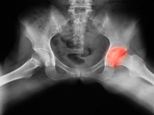
Osteonecrosis of the hip is a gradual disorder that can cause pain when the blood supply to the bone is disrupted. It is estimated that more than 20,000 people in the United States are admitted to the hospital each year for hip osteonecrosis treatment. The condition occurs in individuals of all ages, races, or genders.
What are the causes and risk factors of hip osteonecrosis?
The term osteonecrosis is defined as the death of bone tissue and can cause severe pain and disability. Bone cell death occurs when the blood supply that is responsible for bone nourishment and support is interrupted, which can stem from a variety of causes. The following are some of the most common hip osteonecrosis causes:
- Traumatic injury: Any type of significant injury to the bone leading to bone fractures may interrupt blood supply. Osteonecrosis can occur due to an injury caused by playing contact sports, due to an unforeseen accident, or simply by a joint dislocation that interrupts blood flow.
- Perthes disease: A rare childhood condition affecting the hip and characterized by temporary interruption of blood flow to the rounded head of the thighbone (femur).
- Peripheral vascular disease (PVD): A blood circulation disorder caused by narrowing blood vessels found outside the heart and brain, most commonly occurring in the blood vessels of the lower extremity.
- Slipped capital femoral epiphysis: A condition seen in adolescents that is caused by weakness of the growth plate, causing the femur head to slip backward.
- Sickle cell anemia: An inherited blood cell disorder that is characterized by abnormally shaped hemoglobin that takes the form of a sickle. These sickled cells are fragile and prone to rupture and do not function as normal red blood cells.
- Systemic lupus erythematosus (SLE or lupus): An autoimmune condition that causes the body’s own immune system to mistakenly attack healthy tissue in many parts of the body.
- Decompression sickness: Also known as diver’s disease or the bends. This condition is caused by the formation of gas within the body. The condition is commonly known to manifest when ascending too quickly while scuba diving. The abnormal formation of nitrogen bubbles within the blood can lead to an arterial gas embolism.
- Radiation therapy
- Excess alcohol intake
It is important to note that a risk factor only increases the chances of a particular condition developing. It does not necessarily mean they will definitely get it. Risk factors for the development of hip osteonecrosis include the following:
- Excess consumption of alcohol
- Hip dislocation or fracture
- Systemic lupus erythematosus
- Decompression sickness
- Sickle cell anemia
- Gaucher’s disease
- Crohn’s disease
- Arterial embolism
- Thrombosis
- Vasculitis
- Prolonged consumption of a steroidal medication
What are the signs and symptoms of osteonecrosis of the hip?
Hip osteonecrosis symptoms are commonly hallmarked by a dull ache or throbbing pain in the going or buttock area, during the early stages of the disease. As the condition progresses, movement will become more difficult as it affected patients will have trouble standing up on the own and putting weight on the hip joint. Moving the hip will also elicit pain. Patients will often present with an abnormal way of walking, known as Trendelenburg gait.
How to diagnose hip osteonecrosis?
Hip osteonecrosis diagnosis will depend on the specific classification (stage 0 to IV) of the condition and is dependent on X-ray, MRI, and bone scan appearance. Additional tests may include a core biopsy and venography.
Other factors include the general appearance of the affected region, bone position, estimating percentage volume of the head involved (axial) and percentage weight bearing surface involved (coronal), coexisting osteoarthritis or secondary degenerative change, the presence of joint effusion, and the presence of a potentially unstable osteochondral fragment. Additionally, laboratory tests may be done to identify a potential cause of hip osteonecrosis.
What are the treatment options for osteonecrosis of the hip?
A combination of non-surgical and surgical methods for hip osteonecrosis treatment are often implemented in affected patients, and it will depend on the overall assessment the patient’s particular condition.
- Non-surgical methods include:
- The use of ice to reduce pain and swelling
- Non-steroidal anti-inflammatory medication (e.g. Ibuprofen and Naproxen)
- Corticosteroid injections
- Physical therapy to help restore strength and flexibility to muscles
Surgical methods include:
- Partial hip replacement: Involves removal of one part of the hip joint and is often recommended if the disorder is confined to a certain area of the hip. This area of the hip is replaced with a prosthetic implant.
- Total hip arthroplasty: Involves complete removal of cartilage within the hip joint, which is then replaced by a metal and plastic prosthetic implant. This procedure is often recommended if the disorder affects the entire hip.
- Cartilage grafting: A procedure that replaces damaged hip cartilage.
- Core decompression: Often done in early stages of hip osteonecrosis and characterized by decreasing the pressure within the bone by removing the part of the bone causing abnormal pressure.
Prevention and prognosis of hip osteonecrosis
The following are some general tips for hip osteonecrosis prevention:
- Avoid excessive alcohol consumption
- Wear safety equipment when playing high-risk sports such as football
- Properly manage chronic condition such as SLE, vasculitis, and Crohn’s disease
- Adhere to scuba diving rule for ascension and wear proper scuba diving equipment
- Only take steroidal medication as prescribed
- Maintain a low cholesterol diet
- Avoid or promptly treat blood clots that occur within blood vessels
If recognized in its early stages, followed by promptly implemented treatment measures, hip osteonecrosis prognosis is relatively favorable. However, the extent of bone damage will significantly affect patient outcomes.
Also Read: 16 reasons for pain above the left hip